3D CBCT Imaging in Modern Endodontic Diagnostics
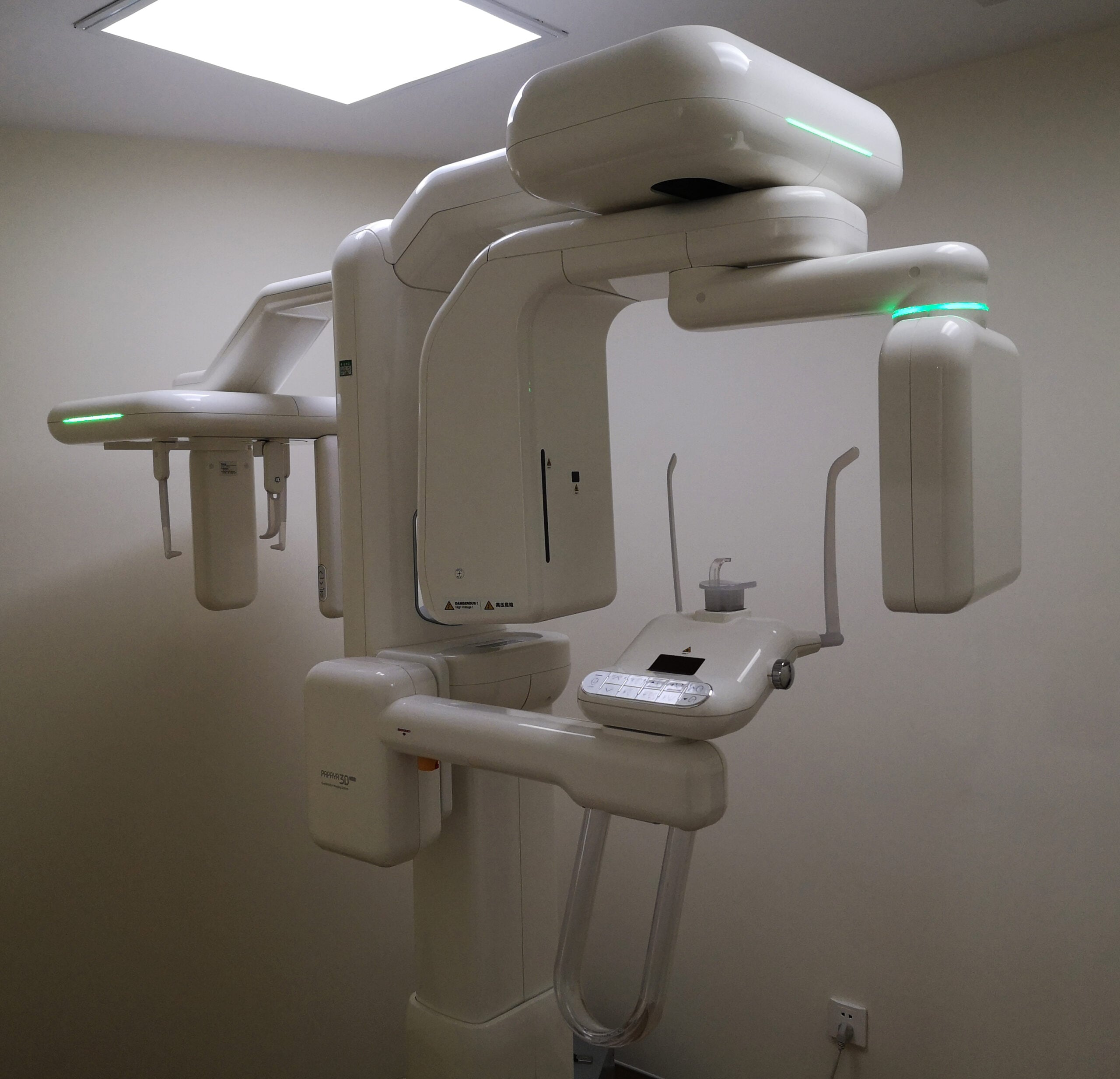
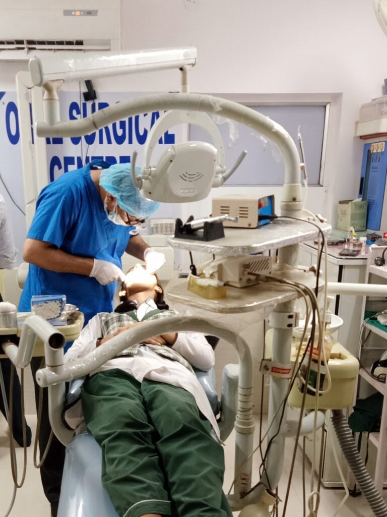
When it comes to endodontic care, seeing clearly is everything. At Anodyne Endodontics in Bloomingdale, IL, we use state-of-the-art 3D Cone Beam Computed Tomography (CBCT) imaging to diagnose and plan treatment with a level of precision that traditional 2D X-rays simply cannot provide.
What Is CBCT Imaging?
3D CBCT imaging creates a three-dimensional model of your teeth, jawbone, nerve pathways, and surrounding structures. Unlike conventional dental X-rays, which produce flat, two-dimensional images, CBCT allows Dr. Zainab Aziz to visualize the full anatomy of your tooth root system from every angle. This technology is particularly valuable in endodontics, where hidden canals, fractures, and infections can lurk in areas invisible to standard radiography.
Why 3D Imaging Changes Root Canal Outcomes
The anatomy of tooth roots is far more complex than most patients realize. A single molar may have three, four, or even five root canals — and variations are common. Studies show that up to 50% of missed canals in failed root canal cases could have been detected with 3D imaging.
At Anodyne, CBCT imaging helps us identify all root canals including curved or unusually located ones, detect cracks before they cause irreversible damage, measure the exact length and curvature of each root for precise instrumentation, locate hidden infections that appear subtle or absent on 2D X-rays, and plan surgical approaches for endodontic microsurgery with millimeter accuracy.
Patient Safety
Modern CBCT units deliver a fraction of the radiation of medical CT scanners. Dr. Aziz follows the ALARA principle (As Low As Reasonably Achievable), using 3D imaging only when it provides clear clinical benefit.
Serving the Western Suburbs
Patients from Bloomingdale, Wheaton, Glen Ellyn, Carol Stream, and Bartlett trust Anodyne Endodontics for the most advanced diagnostic technology available. Call us at 659-ANO-DYNE or schedule a consultation online.