
CBCT Imaging Root Canals: Precision Endodontics Guide
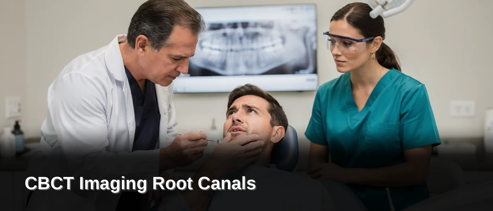
CBCT-guided endodontics uses cone beam computed tomography, a twenty-second 3D scan, to map your tooth’s canals, roots, and surrounding bone with far greater detail than standard X-rays. Dr. Zainab Aziz at Anodyne Endodontics relies on this technology to pinpoint infection sources precisely, guiding treatment that protects healthy tissue and preserves your natural tooth.
Key Takeaways
- CBCT captures 3D dental images in twenty seconds, revealing tooth roots and surrounding bone structure clearly.
- 3D imaging detects fractures, missed canals, and infections that traditional 2D imaging fails to identify.
- CBCT provides superior diagnostic information about dental anatomy and periapical pathologies compared to conventional radiographs.
- Advanced 3D imaging enables precise root canal treatment planning and execution for complex endodontic cases.
What Makes Tooth Pain Hard To Diagnose?
Pain in one tooth often traces back to a completely different tooth, a cracked root, or a hidden canal that never shows up on a apartment X-ray. Your nerves overlap and refer pain across the jaw. The spot that hurts isn’t always the spot that’s actually infected. Guesswork costs you time, and time lets infection spread deeper into the bone.
Two-dimensional X-rays flatten a three-dimensional problem. Clinical literature confirms that 3D imaging is diagnostically superior to traditional apartment, two-dimensional dental X-rays. It captures depth, angle, and hidden canal anatomy a 2D image simply can’t show. At Anodyne Endodontics, your diagnosis relies on CBCT imaging for root canals alongside a dental operating microscope and GentleWave technology, giving your endodontist a complete picture before any drill touches your tooth.
Why can’t a regular dentist always find the source of the pain?
General dentists see a huge range of conditions every day, from cavities to cleanings to crowns. Endodontists complete years of additional training focused entirely on the inside of the tooth, sharpening exactly the skill needed to read complex root anatomy. That specialist-level training, built on two to three additional years beyond dental school, exists specifically to save your natural tooth rather than remove it.
What does 3D imaging actually reveal that a standard X-ray misses?
A CBCT scan builds a rotating 3D model of your tooth, root, and surrounding bone. Your endodontist uses that model for cone beam CT treatment planning, spotting fractures, extra canals, or infection pockets a apartment image hides. This precision also supports 3D guided endodontics, so treatment targets the actual problem instead of a best guess.
How Does CBCT Imaging Root Canals Work?
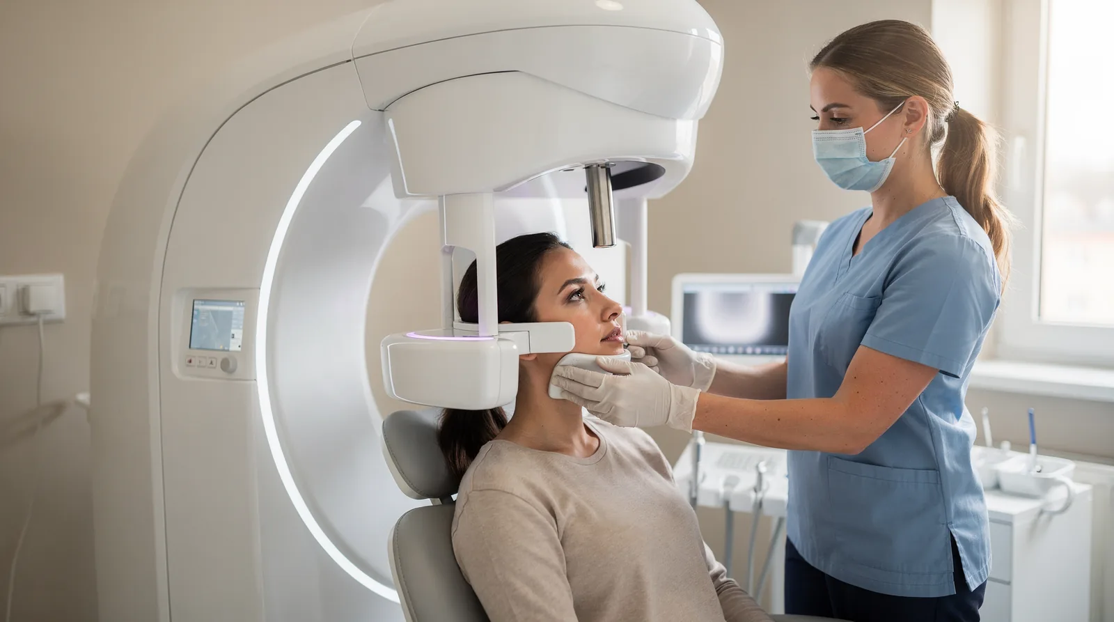
A cone beam scanner captures a full 3D map of your tooth roots, jawbone, and surrounding structures in about twenty seconds. That speed matters: you get precise diagnostic detail without a lengthy appointment or extra time in the chair. Instead of a apartment, two-dimensional picture, your endodontist sees your tooth’s anatomy from every angle before a single instrument touches it.
During the scan, a sensor circles your head in a motion you may recognize from a panoramic X-ray, the kind most dental offices already use. You sit still for a short rotation, and the machine does the rest. No injections, no extended exposure, no guesswork about what’s happening beneath the surface.
Cone beam CT treatment planning turns that scan into a roadmap. Your endodontist reviews the 3D images to locate hidden canals, curved roots, or areas of infection that standard X-rays often miss. This groundwork is where 3D guided endodontics begins: treatment planned with anatomical accuracy rather than approximation.
Why does 3D imaging matter for your root canal outcome?
Hidden canals and unusual root shapes cause many retreatment cases down the road. CBCT imaging root canals gives your endodontist a clearer view upfront, reducing the chance that something gets missed the first time.
What happens after the scan?
Your endodontist combines the CBCT data with an operating microscope and GentleWave irrigation technology to deliver treatment with tighter precision and greater comfort. At Anodyne Endodontics, board-certified endodontist Dr. Zainab Aziz leads the Bloomingdale office, giving patients across the western suburbs access to this microscopic, image-guided care close to home.
Why Choose 3D Guided Endodontics Over Apartment X-Rays?
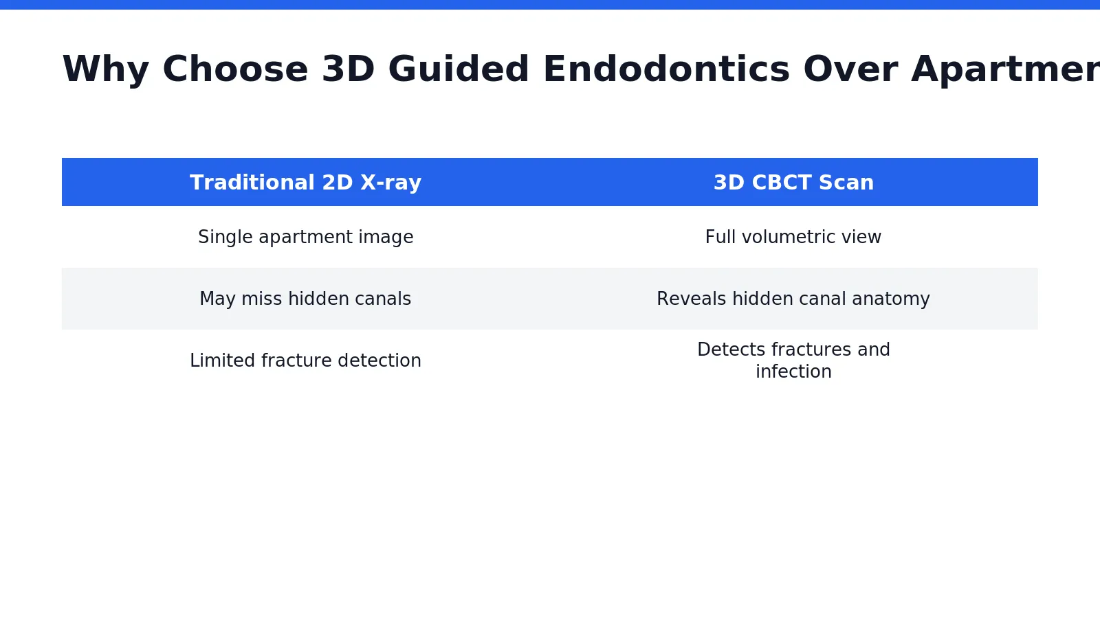
A three-dimensional scan simply shows your dentist more than a apartment X-ray ever could. Where traditional imaging flattens your tooth into a single plane, CBCT imaging root canals captures depth, angle, and structure all at once, giving your specialist a complete map of what’s happening beneath the surface.
That difference matters when pain won’t explain itself. Standard 2D X-rays miss fractures hiding along a root, extra canals tucked out of view, or infection pockets that haven’t shown up on a routine film yet. A cone beam scan catches these details. It does so with meaningfully less radiation than a full medical CT scan. An important reassurance if you’re wary of imaging in general.
Does a dental specialist read these scans differently than a general dentist?
Yes, and the distinction matters for your outcome. An endodontist spends years of additional training beyond dental school focused entirely on the internal anatomy of the tooth. That depth of study means your specialist isn’t just looking at a scan. They’re interpreting it with a trained eye toward saving the natural tooth whenever possible.
What does this mean for your treatment plan?
3D guided endodontics turns imaging into a roadmap rather than a snapshot. Instead of guessing at canal locations or root curvature, your specialist uses cone beam CT treatment planning to chart the exact path before ever picking up an instrument.
Better information up front means fewer surprises mid-procedure — and a plan built specifically around your tooth.
How Does Cone Beam CT Treatment Planning Protect Your Tooth?
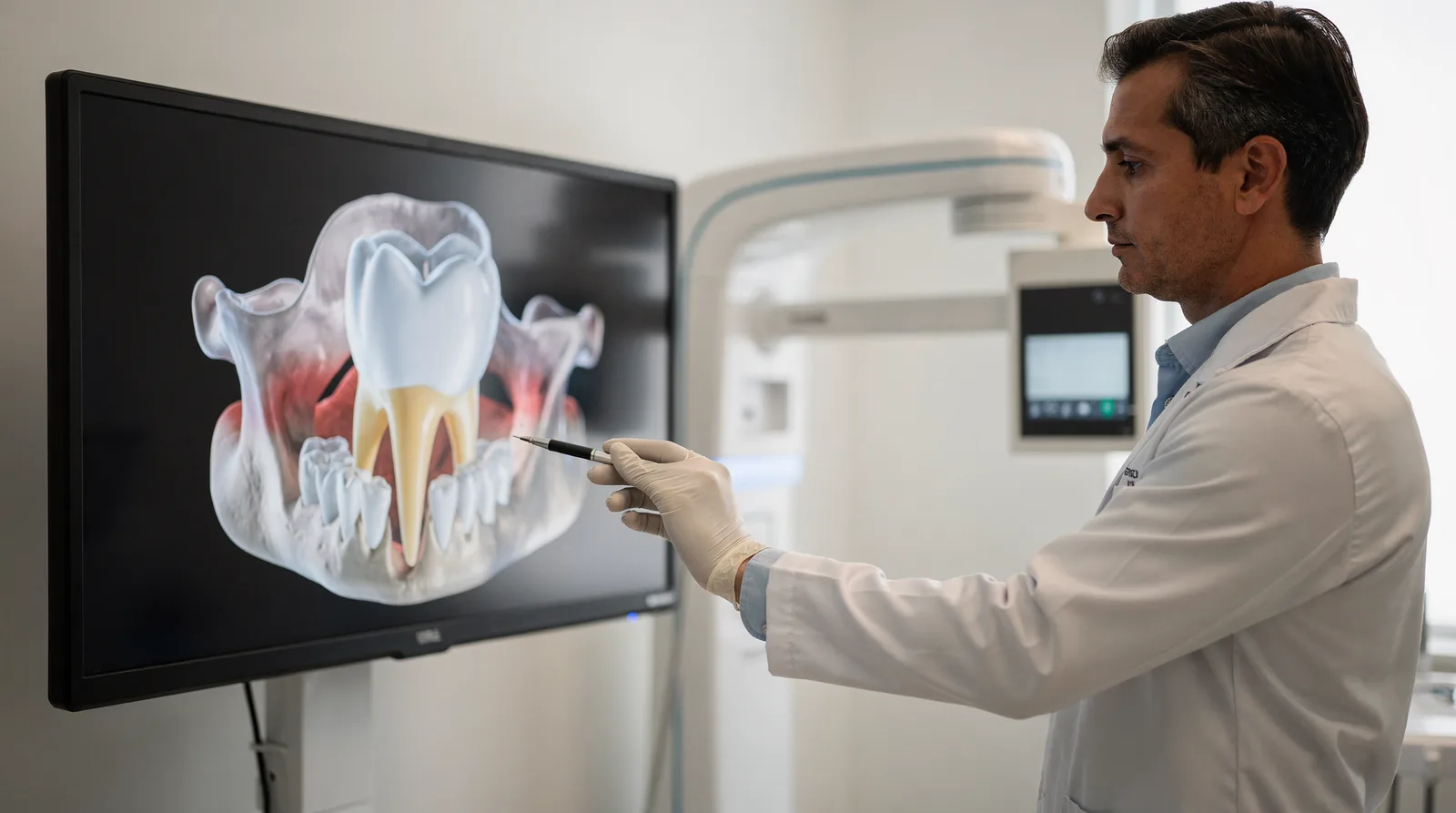
Cone beam CT treatment planning protects your tooth by mapping its internal anatomy in three dimensions before any drilling begins. Instead of guessing at canal shape or root curvature, your endodontist sees the actual terrain first. That single difference changes how conservatively — and how accurately — your treatment unfolds.
A detailed 3D scan reveals exactly which tooth structures are affected and why. That clarity lets your endodontist work with precision instead of exploration, removing only what needs to go and preserving as much of your natural tooth as possible. Less guesswork means less unnecessary drilling into healthy dentin.
What role does CBCT play in guided procedures?
CBCT imaging root canals function as the foundation for 3D guided endodontics, giving your treatment plan a precise scaffold to follow. This imaging is also strongly recommended before your endodontist uses advanced irrigation systems designed to clean canals more thoroughly. Skipping this step leaves those advanced tools working with incomplete information about your tooth’s actual shape.
Why does 3D imaging matter for your specific case?
Every root system is different, and apartment, two-dimensional X-rays can hide extra canals or subtle fractures. Three-dimensional scanning closes that gap.
At Anodyne Endodontics, this technology pairs with a dental operating microscope and GentleWave irrigation to map your treatment before a single canal is touched. For Bloomingdale, IL patients and neighboring DuPage County communities, that combination means:
- Fewer surprises once treatment starts
- Reduced risk of missed canals or undetected fractures
- Treatment focused on saving your natural tooth structure
Losing that upfront clarity means treating blind — a risk your tooth shouldn’t have to carry.
What Happens During Your Bloomingdale CBCT Visit?
Your visit centers on one goal: giving your endodontist a complete, three-dimensional picture of the tooth causing you trouble. Anodyne Endodontics sits in Bloomingdale, IL, putting advanced 3D guided endodontics within easy reach for patients across the western suburbs. You won’t need to travel into the city or navigate a large, impersonal facility to get this level of diagnostic detail.
A compact clinical team makes the difference here. With five staff members total, your scan and consultation move at an unhurried pace, and someone reviews your images with you rather than rushing to the next chart. Under Dr. Zainab Aziz, a board-certified endodontist, that review translates directly into microscopic, image-guided root canal therapy. Nearby communities gain specialist-level care without a long drive.
Why would your endodontist order a CBCT scan before treatment?
Cone beam CT treatment planning is one of the most common reasons a scan gets ordered before complex root canal cases. If your tooth has curved canals, prior treatment, or unclear symptoms on a standard X-ray, a 3D scan helps map the anatomy before any instrument enters the tooth.
What does the scan itself involve?
You sit while a rotating scanner captures your tooth and surrounding bone from multiple angles. The resulting images let your endodontist plan each step with precision, reducing guesswork and protecting healthy tooth structure during treatment.
FAQ
How long does a CBCT scan take?
A cone beam scanner captures a full 3D map of your tooth roots, jawbone, and surrounding structures in about twenty seconds, giving Dr. Aziz precise diagnostic detail without a lengthy appointment.
What can CBCT imaging detect that regular X-rays miss?
CBCT reveals fractures, missed canals, and infections that traditional 2D imaging fails to identify. It captures depth, angle, and hidden canal anatomy a apartment image cannot show.
What technology does Anodyne Endodontics use alongside CBCT?
Dr. Aziz combines CBCT imaging with a dental operating microscope. GentleWave technology, giving your endodontist a complete picture of your tooth before any drill touches it.
Conclusion
In closing, CBCT-guided endodontics represents a fundamental shift in how we diagnose, plan, and execute root canal therapy with precision and confidence. Three-dimensional imaging eliminates diagnostic uncertainty, allowing us to identify complex anatomy, locate calcified canals, and detect periapical pathology that conventional radiographs miss. By integrating this advanced technology into our clinical workflow, we deliver superior outcomes while minimizing procedural time and tissue trauma. For patients seeking the highest standard of endodontic care, CBCT guidance transforms root canal treatment from a necessary intervention into a predictable, comfortable healing experience.