Understanding Calcified Canals: Why Modern Imaging and Advanced Techniques Preserve Difficult Root Canals
By Zainab Aziz, Anodyne Endodontics
Calcified canals are a common challenge in endodontics, presenting significant obstacles during root canal treatments. As dental anatomy varies greatly among individuals, the presence of calcifications can complicate access to the pulp and make necessary procedures more arduous. In this article, we will explore the nature of calcified canals, the innovative imaging techniques available to diagnose them, and the advanced treatments that can manage their complexity effectively. By understanding these concepts, both dental professionals and patients can appreciate the advancements that improve treatment outcomes.
The discussion will encompass various aspects, starting with a definition of calcified canals, delving into the enhancements provided by Cone Beam Computed Tomography (CBCT) imaging, and concluding with the advanced techniques used in their management. Finally, guidance will be provided on when general dentists should consider referring patients to an endodontist.
What Are Calcified Canals and How Do They Affect Root Canal Treatment?
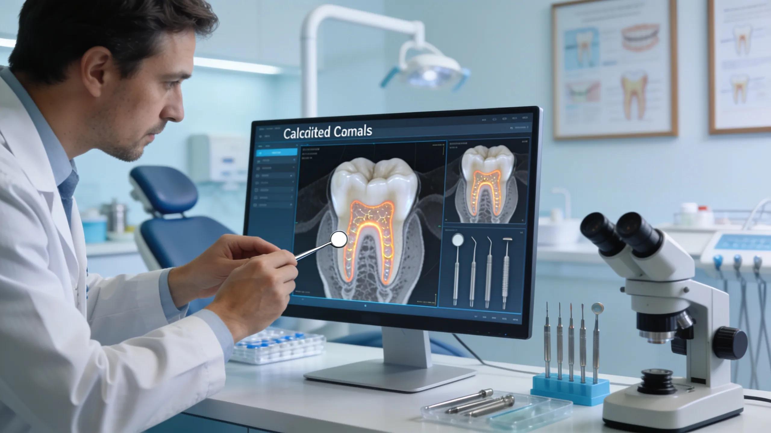
Calcified canals refer to the condition where the root canal space becomes narrowed or obstructed due to the deposition of mineralized tissue, impeding access to the pulp. This presents challenges during root canal therapy, as procedures may require specialized techniques to navigate through narrowed pathways. The presence of calcifications can lead to missed canals or incomplete cleaning, ultimately impacting success rates in managing dental pulpal issues. Recognizing and addressing these challenges is critical for achieving effective treatment outcomes.
Understanding the implications of calcified canals highlights the necessity for advanced tools and techniques in endodontics. Accurate diagnosis and treatment planning are essential to mitigate the risks associated with these abnormalities.
To further understand how these mineralized deposits alter tooth structure and complicate clinical procedures, researchers have explored the specific impacts of calcific metamorphosis on canal negotiation.
Understanding Calcific Metamorphosis and Canal Negotiation
One such complication is Calcific Metamorphosis (CM) which can complicate entry in to the root canal system in the future due to difficulty in accessing the canal. It is the Calcific deposits of the pulp chamber that causes the darker hue, translucency loss and the yellowish appearance of the tooth’s crown. Locating the canal and negotiating it to full working length may lead to iatrogenic errors such as fractured instrument and perforation. Calcified canal and negotiation-A review, 2018
How Does CBCT Imaging Enhance Diagnosis and Treatment of Calcified Canals?
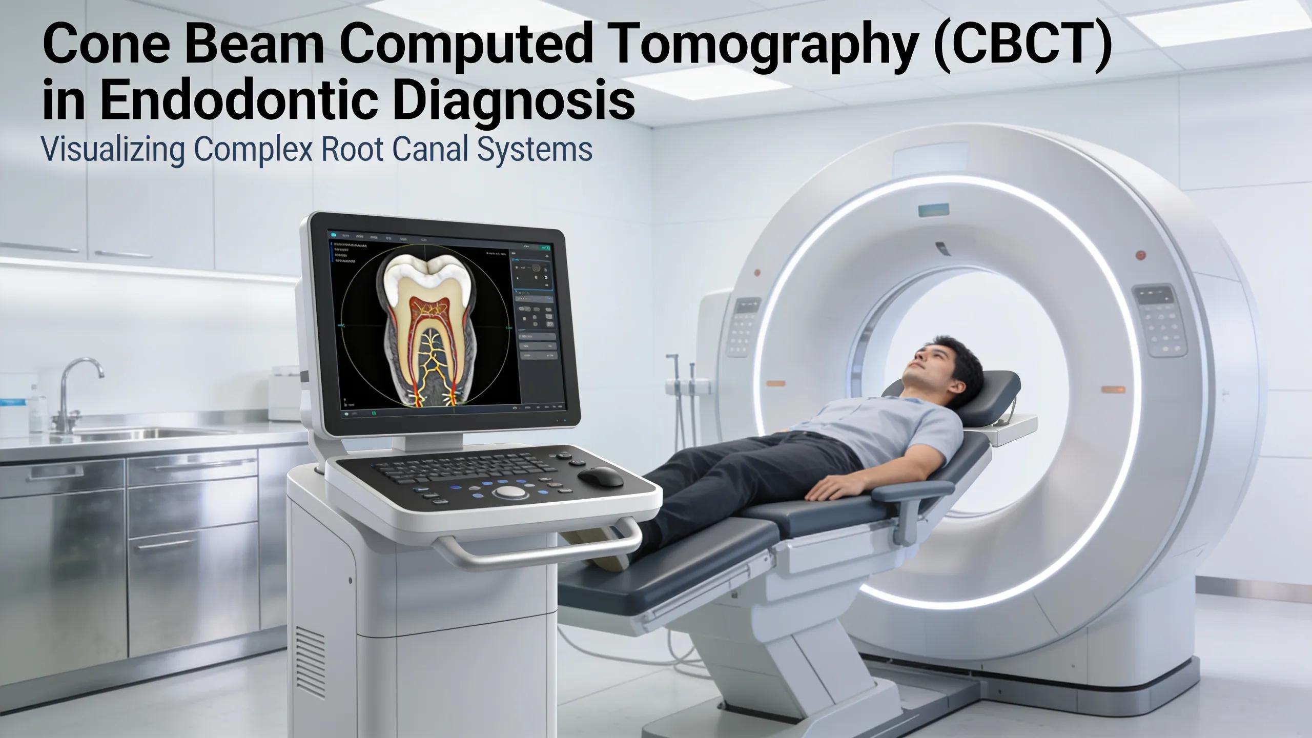
Cone Beam Computed Tomography (CBCT) imaging enhances the diagnosis and treatment of calcified canals by providing three-dimensional representations of dental anatomy. This enhanced visualization of complex anatomical structures plays a crucial role in identifying calcified canals. With a significant reduction in missed canals, CBCT allows practitioners to accurately assess the topography of root canal systems before proceeding with treatment.
This imaging technique improves overall diagnostic accuracy, enabling better treatment planning and execution for challenging cases. When evaluating complex endodontic cases, it becomes essential to employ technologies like CBCT to ensure thorough and effective treatment approaches.
Anodyne Endodontics is dedicated to patient education and utilizing advanced technologies, allowing for optimized diagnosis and treatment plans for calcified canals.
Integrating modern three-dimensional imaging with innovative intraoperative markers has revolutionized how clinicians plan and execute interventions for severely obstructed roots.
CBCT and Radiopaque Markers in Calcified Root Canals
The aim of this study was to describe a new strategy, consisting of the use of cone-beam computed tomography (CBCT) in the planning and intraoperative stages of root canal treatment (RCT), associated with the use of radiopaque gutta-percha markers, as an auxiliary tool in the location of severely calcified root canals.
Root canal treatment of severely calcified teeth with use of cone-beam computed tomography as an intraoperative resource, IBF Petean, 2022
What Advanced Techniques Are Used to Manage and Treat Calcified Root Canals?
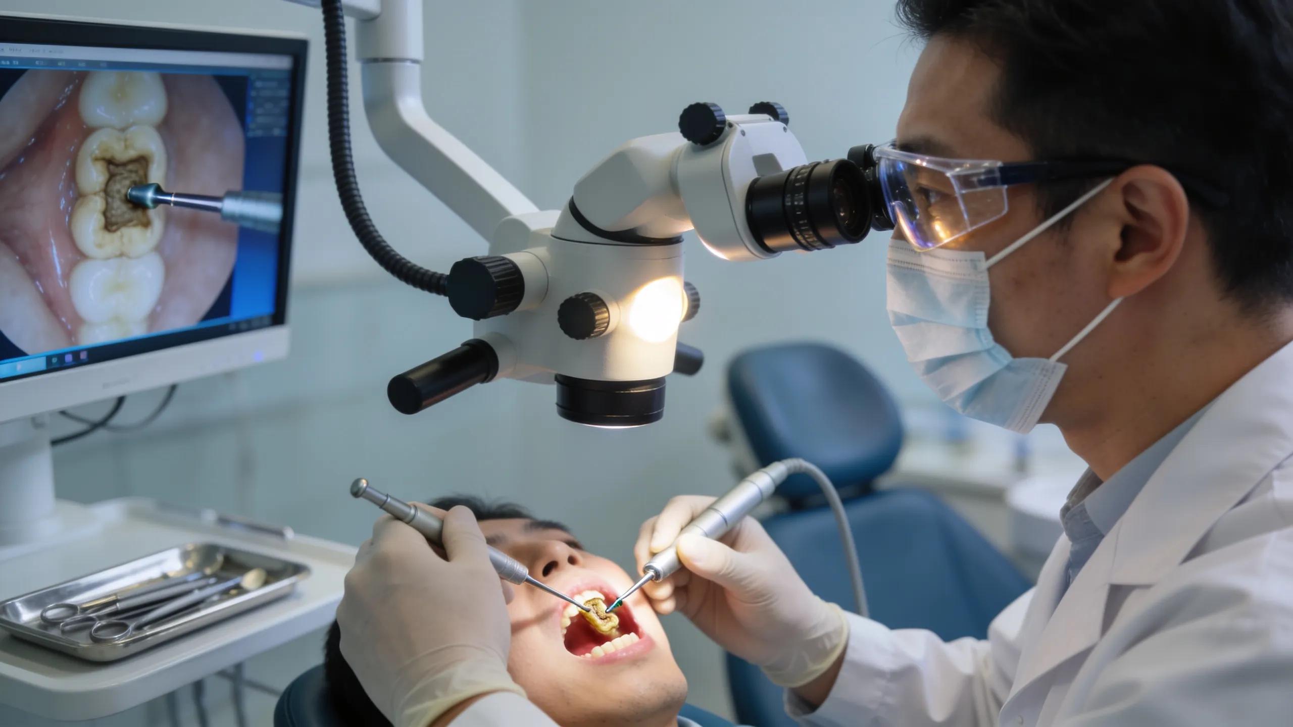
Managing calcified root canals necessitates the application of advanced techniques and tools, certifying the best possible outcomes. Dental professionals implement various methods, adapting to the specific challenges each case poses. Key technologies utilized include:
- CBCT Imaging: Facilitates detailed visualization of the root canals, crucial for accurate treatment planning.
- Dental Operating Microscope: Enhances visibility and precision during endodontic procedures, allowing for more effective interventions.
- GentleWave System: Utilizes advanced fluid dynamics to optimize cleaning and disinfection of root canal systems.
These advanced approaches enhance procedural efficacy and improve patient outcomes by minimizing the risk of complications associated with calcified canals.
Comparison of Techniques for Treating Calcified Root Canals
This comparison underscores how innovative techniques foster superior treatment outcomes for calcified canals.
When Should General Dentists Refer Patients With Calcified Canals to an Endodontist?
The referral of patients to an endodontist becomes crucial when certain clinical indicators present themselves. General dentists should consider referring in situations such as:
- Persistent Symptoms: Ongoing pain or discomfort despite initial treatment efforts suggests deeper issues.
- Identifying Complex Anatomical Features: Difficulty visualizing or managing complex root canal morphology indicates a need for specialized expertise.
- Failed Endodontic Treatment: Cases of retreatment or complications following prior procedures warrant referral to ensure optimal outcomes.
These referral criteria enable general practitioners to leverage the specialized skills of endodontists for managing complex cases effectively. Ensuring patients receive timely and appropriate referrals can significantly enhance their treatment experiences and outcomes.